micro-aneurysm
Cellular flow in 3D reconstructed retinal micro-aneurysm
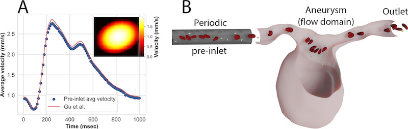
Blood flow within the vasculature of the retina has been found to influence the progression of diabetic retinopathy. In this research cell resolved blood flow simulations are used to study the pulsatile flow of whole blood through a segmented retinal microaneurysm. Images were collected using adaptive optics optical coherence tomography of the retina of a patient with diabetic retinopathy, and a sidewall (sacciform) microaneurysm was segmented from the volumetric data. The original microaneurysm neck width was varied to produce two additional aneurysm geometries in order to probe the influence of neck width on the transport of red blood cells and platelets into the aneurysm. Red blood cell membrane stiffness was also increased to resolve the impact of rigid red blood cells, as a result of diabetes, in blood flow. Wall shear stress and wall shear stress gradients were calculated throughout the aneurysm domains, and the quantification of the influence of the red blood cells is presented. Average wall shear stress and wall shear stress gradients increased due to the increase of red blood cell membrane stiffness. Stiffened red blood cells were also found to induce higher local wall shear stress and wall shear stress gradients as they passed through the leading and draining parental vessels. Stiffened red blood cells were found to penetrate the aneurysm sac more than healthy red blood cells, as well as decreasing the margination of platelets to the vessel walls of the parental vessel, which caused a decrease in platelet penetration into the aneurysm sac.
References
2022
- The effect of stiffened diabetic red blood cells on wall shear stress in a reconstructed 3D microaneurysmComputer Methods in Biomechanics and Biomedical Engineering, 2022